


Key Points:
- Measures of intelligence decline as age increases in epilepsy patients.
- Neuron-support cells, called astroglia cells, increased as measures of intelligence decreased.
- While preliminary, the findings suggest that maintaining astroglia cell health with age could potentially counteract intelligence loss.
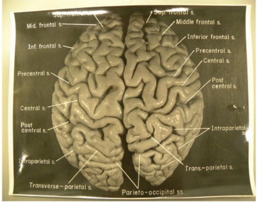
In 1985, scientists found that Albert Einstein’s brain contained more glial cells relative to neurons. Glial cells are a class of brain cells that help nourish neurons, insulate nerve fibers, regulate inflammation, and maintain cell-to-cell communication.
Now, USC researchers have found that glial cells, specifically astroglia, may support the maintenance of intelligence with age. In a new study published in Frontiers in Cellular Neuroscience, older epilepsy patients had more abnormal immature astroglia than their younger counterparts. Furthermore, higher numbers of these cells corresponded with lower scores on tests of fluid intelligence—the capacity to reason quickly, think abstractly, and solve problems independent of knowledge.
Understanding Accelerated Brain Aging
In aging biology, aging does not necessarily equate to the number of years lived (chronological age). As we age, cellular damage accumulates despite the body’s repair mechanisms, impairing tissues and organs and increasing the risk of disease and death. When this deterioration occurs earlier than expected, it is called accelerated aging. Our organs also age at different rates, and their biological ages can help predict mortality risk, highlighting how unevenly the body ages.
Epilepsy Accelerates Brain Aging
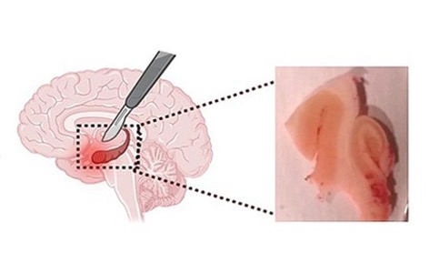
The brain, for example, can show signs of accelerated aging in people with epilepsy, specifically mesial temporal lobe epilepsy. Studying these patients may therefore offer valuable insights into how the brain ages. Moreover, some patients whose seizures do not respond to medication undergo surgery to remove affected brain tissue, which may include part or all of one hippocampus—a structure involved in forming and consolidating memories. When donated for research, this tissue offers a rare opportunity to examine the cellular features of brain aging directly.
Fluid Intelligence and Age
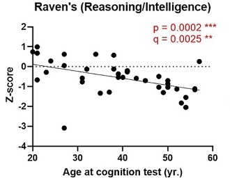
The researchers analyzed pre-surgical cognitive tests from 40 patients, whose ages ranged from 20 to 58. The assessments measured abilities ranging from attention and language to memory and abstract reasoning. Scores were adjusted for education to account for differences in schooling.
Older patients performed worse in three areas: fluid intelligence, visuospatial reconstruction, and visuospatial memory. These abilities help us solve unfamiliar problems, reproduce visual arrangements, and remember what we have seen. Unlike knowledge accumulated through experience, fluid intelligence involves recognizing patterns and reasoning through situations without relying on previously learned answers.
More Immature Astroglia, Lower Reasoning Scores
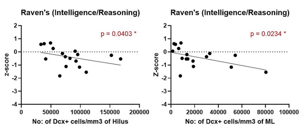
The researchers next examined hippocampal tissue by counting abnormal, immature astroglia carrying doublecortin (Dcx), a protein used to identify this cell population. They found higher-than-normal immature astroglia counts in two hippocampal regions, which corresponded to lower fluid intelligence scores. Notably, the same relationship did not appear for visuospatial reconstruction or memory, pointing toward a more specific connection with reasoning.
The Study’s Limits
Because the study was observational, it cannot establish whether immature astroglia contribute to cognitive decline or merely accompany other brain changes. Moreover, immature astroglia have not been observed in healthy older adults. The study also compared patients of different ages rather than tracking changes within individuals, and its findings may not extend to people without epilepsy. Additionally, the associations did not withstand more robust statistical correction (multiple comparisons or adjustment for age). It follows that the results remain preliminary. However, maintaining glial cell health with age may still potentially counteract intelligence loss and protect against cognitive decline.
Maintaining the Brain’s Support System
Physical activity offers one practical avenue for maintaining glial cell health with age. Human research has associated greater physical activity with better cognition and the improved health of microglia, the brain’s resident immune cells that modulate inflammation. Experimental research also suggests that exercise can influence astroglia function and interactions with neurons.
Another avenue involves cellular senescence, a dysfunctional state that can develop with aging. A recent study showed that senescent astroglia lose some of their ability to support communication between neurons. Thus, reducing senescent cells could potentially mitigate this deficit. With that said, a multiple-component nutraceutical, Restorin, was designed to reduce cellular senescence and other detrimental age-related targets. However, while these targets provide a rationale for investigating possible effects on aging glia, they do not establish that the product maintains these cells in people.
 Comments