


Key Points:
- Chronically stressed mice have more senescent cells and lower L-serine levels in their brain than normal.
- Giving stressed mice L-serine, or a senolytic called DQ (dasatinib and quercetin), counters stress-induced senescent cells, anxiety-like behavior, and metabolic dysregulation, including high blood glucose levels.
- Both interventions help rebalance the nervous system of the stressed mice by reducing “fight-or-flight” activation and increasing “rest-and-digest” activation.
Chronic stress isn’t purely psychological, but can wreak havoc on the body. In a new study published in Cell Metabolism, researchers in China found that chronic stress induced insulin resistance in mice. Insulin resistance is implicated in several chronic conditions, including type 2 diabetes, cardiovascular disease, and Alzheimer’s disease.
The researchers also reported that L-serine reduced stress-associated insulin resistance by acting on senescent cells in the amygdala, a brain region involved in processing emotions such as fear. A senolytic combination of dasatinib and quercetin (DQ), which is designed to selectively eliminate senescent cells, produced similar metabolic benefits.
Stressed mice also developed elevated blood glucose levels, suggesting that prolonged stress may trigger diabetes-like metabolic changes—at least in mice. The findings raise the possibility that clearing senescent cells, or preventing stress from inducing them in the first place, could help protect against the metabolic consequences of chronic stress.
How to Chronically Stress Mice
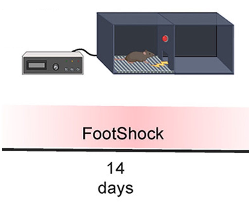
To generate a model for chronic stress, the researchers subjected mice to 14 days of foot shocks. The mice were placed into a chamber with an electrified grid, and their feet were shocked daily. The researchers referred to these mice as foot-shock (FS) mice. The mice showed anxiety-like behavior, as assessed by several tests, in which the FS mice were less likely to explore open areas. Moreover, metabolic measurements suggested that the FS mice had insulin resistance and high blood glucose levels.
Chronic Stress Associated with Senescence and Reduced L-Serine
Chronic stress, such as that from social anxiety or phobias, increases the activity of the amygdala, which tends to activate in response to perceived threats. In light of this brain structure’s role in stress, the researchers measured genes from the amygdala of FS mice. They found elevations in genes associated with senescent cells, which are triggered by cellular stressors like DNA damage or metabolic stress.
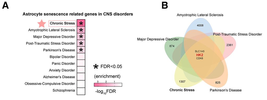
The increase in senescence-associated genes was concentrated in astrocytes—brain support cells that help nourish, protect, and regulate neurons. Notably, the researchers’ analysis suggested that astrocyte senescence-related genes are shared across several neurological and psychiatric conditions, including major depressive disorder, post-traumatic stress disorder (PTSD), and Parkinson’s disease.
They also identified altered activity of hexokinase 2 (HK2) across multiple conditions. Because HK2 helps cells process glucose, this finding may point to a shared link between disrupted glucose metabolism and brain disorders like chronic stress.
Genetically deleting HK2 specifically in mouse amygdala astrocytes largely reproduced the effects seen in FS mice: anxiety-like behavior, elevated blood glucose, and insulin resistance. The researchers then found that astrocyte HK2 deficiency reduced L-serine production.
Astrocytes metabolize glucose not only to meet their own energy needs, but also to produce L-serine, a nonessential amino acid. They then supply L-serine to neurons, where it can be converted into D-serine—a signaling molecule that supports neuronal development, synaptic plasticity, and communication. The researchers showed that astrocyte HK2 deficiency reduced D-serine synthesis in neighboring neurons and altered neuronal complexity.
These findings suggest that senescent astrocytes fail to transfer enough L-serine to neurons, so the neurons cannot produce enough D-serine to function properly. The lack of D-serine and subsequent neuronal dysfunction may explain the anxiety-like behavior of the FS mice. Neuronal dysfunction could also explain the alterations in glucose metabolism. This is because amygdala neurons connect to the body, to organs like the pancreas. The pancreas controls insulin, a hormone that signals cells to take in glucose from the bloodstream.
DQ and L-Serine Supplementation Counter Consequences of Chronic Stress
To explore the effects of alleviating senescence or restoring L-serine, the researchers injected FS mice with DQ or L-serine. While DQ was injected into the bloodstream, L-serine was injected into the brain. Still, both interventions reduced the anxiety-like behavior, high blood glucose, and insulin resistance exhibited by the FS mice.
Similar results were shown in the mice genetically lacking HK2 in their amygdala astrocytes. By connecting electrodes to the amygdala of these mice, the researchers found increased firing rates, indicating an overactive amygdala. Remarkably, both DQ and L-serine calmed the amygdala, returning firing rates to those of unstressed mice.
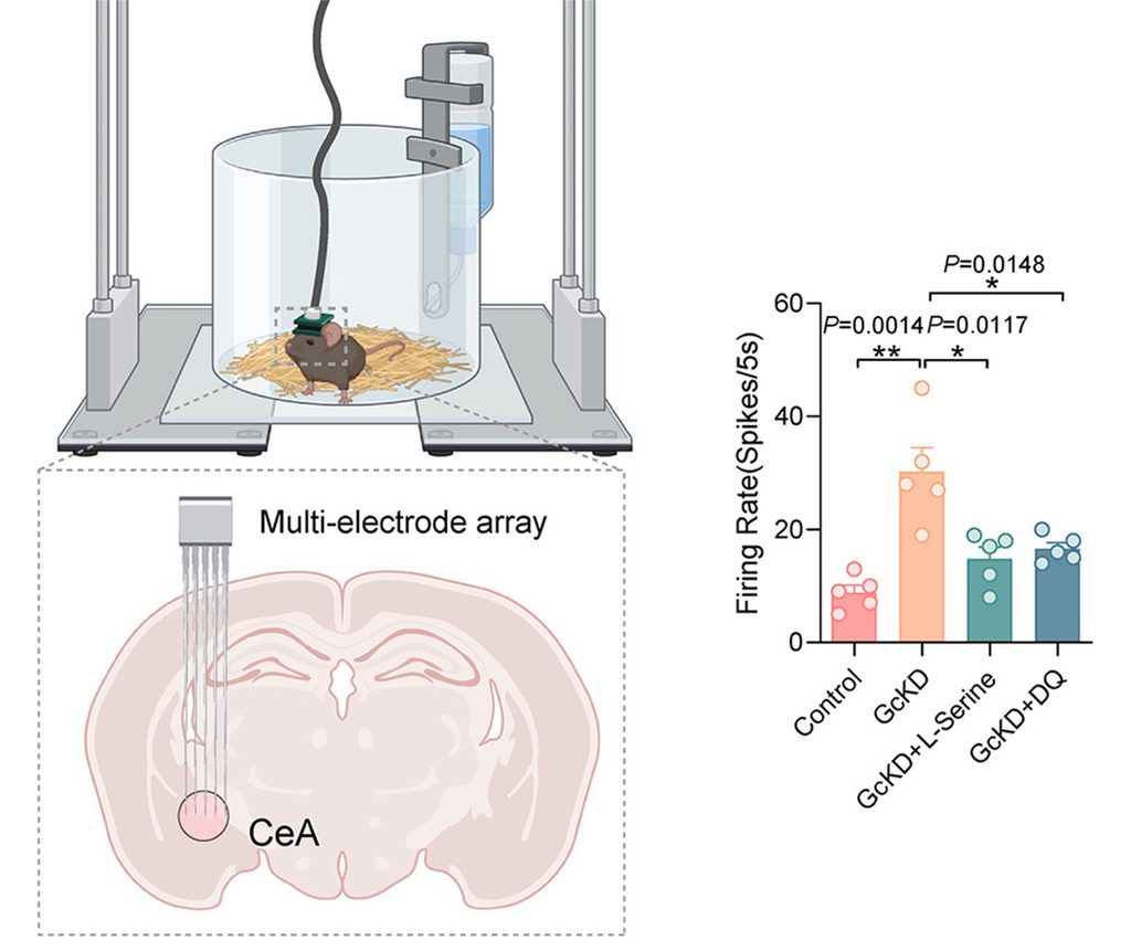
Additionally, in both mouse models, DQ and L-serine reduced markers of senescence in the amygdala. Both interventions also increased L-serine and D-serine levels in the amygdala. Together, these findings suggest that DQ and L-serine supplementation counter stress-induced anxiety, metabolic dysregulation, and senescence.
DQ and L-Serine Help Rebalance Neuronal Control of Pancreas
The pancreas, which releases insulin to help regulate blood glucose, communicates closely with the brain through the autonomic nervous system—the branch of the nervous system that controls involuntary functions. The autonomic nervous system has two main divisions:
- The parasympathetic nervous system, which generally supports “rest-and-digest” functions.
- The sympathetic nervous system, which coordinates the body’s “fight-or-flight” response.
Both divisions send signals to the pancreas and can influence insulin release. Parasympathetic activity generally promotes insulin secretion, particularly around feeding, whereas sympathetic activation during stress can inhibit insulin secretion, contributing to high blood glucose levels.
The researchers found that chronic stress induced an increase in “fight-or-flight” sympathetic nerves surrounding insulin-secreting cells in the pancreas of FS mice. This was accompanied by a reduction in “rest-and-digest” parasympathetic nerves. These findings suggest that stress allows “fight-or-flight” nerves to dominate the control of insulin and blood glucose levels. Supplementing the FS mice with DQ or L-serine rebalanced the nerves surrounding insulin-secreting cells.
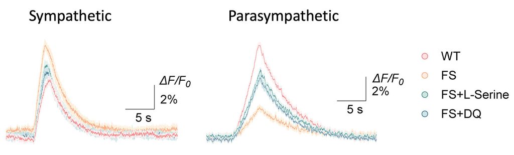
What’s more, to determine the effect of stress in real time, the researchers measured the activation of sympathetic and parasympathetic neurons in the amygdala using a fluorescent dye. They found that FS mice exhibited greater activation in “fight-or-flight” neurons and a reduction in “rest-and-digest” neurons. Remarkably, FS mice treated with DQ or L-serine showed an increase in “rest-and-digest” activation and a reduction in “fight-or-flight” activation. Together, these findings suggest that stress may dysregulate blood glucose levels by turning on the “fight-or-flight” response in the brain, which is connected to the pancreas.
Taking L-Serine or Senolytics to Mitigate the Effects of Chronic Stress
There is not yet convincing evidence that L-serine or DQ supplementation mitigates the pathological effects of chronic stress. However, chronic stress is increasingly recognized as a contributor to insulin resistance and type 2 diabetes risk. In humans, sustained psychosocial stress is associated with poorer glucose regulation and a higher likelihood of developing type 2 diabetes, although the relationship is shaped by many biological and behavioral factors. Biohackers may still wish to try L-serine or DQ to mitigate the effects of stress, based on this study.
 Comments