AGE/DOSE calc
user-icon
Aging & Longevity

Does Fasting Really Trigger Autophagy? New Study Challenges Popular Longevity Belief

A new scientific review questions how much researchers actually know about fasting’s ability to enhance cellular recycling—particularly in humans.

The spherical structure depicts an autophagolysosome.
By Griffin Dean

Key Points:

  • Many studies measure autophagy-related genes and proteins, which cannot independently establish whether autophagy increases.
  • Animal experiments show that prolonged fasting can stimulate autophagy, but human findings remain limited and inconsistent.
  • Researchers still cannot definitively say how long people need to fast to increase autophagy—or which tissues respond.

Fasting is often promoted as a way to remove damaged cellular material and potentially slow aging. One of the proposed mechanisms is autophagy, a recycling process that helps cells dispose of unwanted components and recover nutrients, especially when food is scarce. But does fasting reliably increase autophagy in people? A review published in Biochemical Society Transactions, titled “Increasing autophagy with fasting and diet: separating fact from fiction,” examines the evidence behind this assumption. 

Measuring Autophagy’s Machinery Is Not the Same as Measuring Its Activity

Autophagy allows cells to break down and recycle damaged proteins and other unwanted material, which are packaged into structures called autophagosomes and delivered to lysosomes—the cell’s digestive centers—for breakdown. This process involves many proteins. 
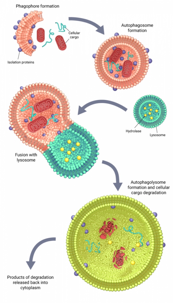
(Image: mbi.nus.edu) Autophagy. A cup-shaped membrane called a phagophore wraps around cellular material until it closes to form an autophagosome. Autophagosomes fuse with lysosomes to form autophagolysosomes, which break down organic material with a low pH (acidic) and enzymes like hydrolases.

Many researchers assess autophagy indirectly by measuring the proteins involved in this process or their messenger RNA (mRNA), the genetic instructions cells use to make those proteins. These measurements can reveal changes in the cell’s recycling machinery, like a snapshot in time. However, they do not confirm whether cellular material has actually been degraded for recycling. Having more instructions for making a protein (mRNA)—or more of the protein itself—does not necessarily mean the machinery is actively breaking down cellular material. 

To more accurately determine whether autophagy has increased, researchers can measure autophagic flux: the process of cellular material moving through the autophagy pathway over time. This distinction is central to interpreting fasting research. Changes in autophagy-related mRNA or protein levels may suggest a response to fasting, but without flux measurements, they cannot confirm that fasting increased autophagy. 

Prolonged Fasting: Strong Animal Evidence, Limited Human Confirmation

Prolonged fasting, defined in the review as at least 24 hours without calories, can robustly stimulate autophagy in animals. Studies have shown, using autophagy markers (proteins and/or mRNA) or autophagic flux measurements, that prolonged fasting can stimulate autophagy in animals. For example, a landmark study showed that 24–72 hours of fasting stimulate autophagy in mice. More recent work using improved techniques showed increased autophagy in liver and muscle, but not the brain, illustrating that tissues respond differently.

Human findings are less conclusive. A three-day fast study showed increased autophagy-related proteins in white blood cells. Muscle studies (Dethlefsen, Vendelbo, Bak) involving 36- or 72-hour fasts also detected changes in signaling or protein abundance, but did not establish increased autophagic flux. Meanwhile, a 26-hour fast study showed that there was little effect on broader autophagy-related gene expression in fat tissue.

One study examined flux after a 24-hour fast but found no increase, despite changes in autophagy-related proteins. Those protein changes appeared only after blood cells were transferred into laboratory dishes, which alters their nutritional environment and complicates interpretation. Overall, the authors conclude that there is no definitive human evidence establishing whether, when, and in which tissues prolonged fasting increases autophagy.

Intermittent Fasting: Responses Depend on the Protocol and Tissue

Intermittent fasting alternates fasting periods with periods when food is available. In a fruit fly study, alternate-day 20-hour fasts increased autophagic turnover and extended lifespan by up to 18%. Disabling essential autophagy genes eliminated that lifespan extension. In a mouse study, alternate-day fasting increased cardiac autophagy during fasting, but not on fed days. 

Human evidence remains sparse. A month-long 17–19 h/day religious fasting regimen increased some autophagy-related mRNA transcripts in blood while decreasing another. A separate regimen involving three non-consecutive weekly 24-hour fasts reduced certain transcripts on non-fasting days in muscle. Neither established increased recycling activity.

Time-Restricted Eating: Meal Timing May Matter

Time-restricted eating, a form of intermittent fasting, confines food intake to a daily window. In a study where mice were given the same total food as controls but limited to two daily two-hour feeding windows, autophagic flux increased immediately after feeding in fat, muscle, and brain tissue. This challenges the simple assumption that eating always suppresses autophagy.

In humans, a six-hour eating window increased an autophagy-related transcript in blood, but flux was not measured. A trial combined a four-hour eating window with substantial (70%) energy restriction on three nonconsecutive days weekly. Participants had higher blood-cell flux than standard-care controls after six months. Because the intervention combined meal timing and reduced calories, their individual contributions remain unclear.

Supporting Autophagy Without Fasting?

Fasting is not the only approach being investigated for its effects on cellular recycling. Supplements may also influence autophagy, although none is an established way to increase it throughout the human body.

Spermidine

In a randomized pilot trial involving 40 adults over 65, supplementation increased autophagic flux in immune cells after two weeks compared with placebo.

Urolithin A

This supplement is being studied for stimulating mitophagy, the selective autophagy of damaged mitochondria. Human trials have reported improvements in some measures of muscle function and changes in mitochondrial and mitophagy-related proteins. However, those markers alone do not establish increased mitochondrial turnover.

Overall, the key question is not whether an intervention changes autophagy-related markers, but whether it increases autophagic flux in relevant tissues—and whether that improves health and longevity. As with fasting, stronger human evidence is needed before these strategies can be considered proven autophagy-based anti-aging interventions.

Source

Singh, S., & Sargeant, T. J. (2026). Increasing autophagy with fasting and diet: separating fact from fiction. Biochemical Society transactions, 54(10), 1497–1511. https://doi.org/10.1042/BST20260632

References

Bak, A. M., Møller, A. B., Vendelbo, M. H., Nielsen, T. S., Viggers, R., Rungby, J., Pedersen, S. B., Lunde Jørgensen, J. O., Jessen, N., & Møller, N. (2016). Differential regulation of lipid and protein metabolism in obese vs. Lean subjects before and after a 72-h fast. American Journal of Physiology-Endocrinology and Metabolism. https://doi.org/E-00464-2015

Bensalem, J., Teong, X. T., Hattersley, K. J., Hein, L. K., Fourrier, C., P. Dang, L. V., Singh, S., Liu, K., Wittert, G. A., Hutchison, A. T., Heilbronn, L. K., & Sargeant, T. J. (2025). Intermittent time-restricted eating may increase autophagic flux in humans: An exploratory analysis. The Journal of Physiology, 603(10), 3019-3032. https://doi.org/10.1113/JP287938

Chaudhary, R., Liu, B., Bensalem, J., Sargeant, T. J., Page, A. J., Wittert, G. A., Hutchison, A. T., & Heilbronn, L. K. (2022). Intermittent fasting activates markers of autophagy in mouse liver, but not muscle from mouse or humans. Nutrition, 101, 111662. https://doi.org/10.1016/j.nut.2022.111662

Defour, M., R. Michielsen, C. J., Afman, L. A., & Kersten, S. (2020). Transcriptomic signature of fasting in human adipose tissue. Physiological Genomics. https://doi.org/PG-00083-2020

Dethlefsen, M. M., Bertholdt, L., Gudiksen, A., Stankiewicz, T., Bangsbo, J., Plomgaard, P., & Pilegaard, H. (2018). Training state and skeletal muscle autophagy in response to 36 h of fasting. Journal of Applied Physiology. https://doi.org/JAPPL-01146-2017

Erlangga, Z., Ghashang, S. K., Hamdan, I., Melk, A., Gutenbrunner, C., & Nugraha, B. (2023). The effect of prolonged intermittent fasting on autophagy, inflammasome and senescence genes expressions: An exploratory study in healthy young males. Human Nutrition & Metabolism, 32, 200189. https://doi.org/10.1016/j.hnm.2023.200189

Godar, R. J., Ma, X., Liu, H., Murphy, J. T., Weinheimer, C. J., Kovacs, A., Crosby, S. D., Saftig, P., & Diwan, A. (2015). Repetitive stimulation of autophagy-lysosome machinery by intermittent fasting preconditions the myocardium to ischemia-reperfusion injury. Autophagy, 11(9), 1537–1560. https://doi.org/10.1080/15548627.2015.1063768

Jamshed, H., Beyl, R. A., Della Manna, D. L., Yang, E. S., Ravussin, E., & Peterson, C. M. (2019). Early Time-Restricted Feeding Improves 24-Hour Glucose Levels and Affects Markers of the Circadian Clock, Aging, and Autophagy in Humans. Nutrients, 11(6), 1234. https://doi.org/10.3390/nu11061234

Kanda, Y., Eguchi, T., Morishita, H., Hama, Y., Abe, M., Sakimura, K., & Mizushima, N. (2026). High basal autophagic activity in the brain revealed by systemic quantitative analysis using GFP-LC3-RFP mice. eLife, 15, Article RP111982. https://doi.org/10.7554/eLife.111982.1

Klionsky, D. J., Abeliovich, H., Agostinis, P., Agrawal, D. K., Aliev, G., Askew, D. S., Baba, M., Baehrecke, E. H., Bahr, B. A., Ballabio, A., Bamber, B. A., Bassham, D. C., Bergamini, E., Bi, X., Biard-Piechaczyk, M., Blum, J. S., Bredesen, D. E., Brodsky, J. L., Brumell, J. H., Brunk, U. T., … Deter, R. L. (2008). Guidelines for the use and interpretation of assays for monitoring autophagy in higher eukaryotes. Autophagy, 4(2), 151–175. https://doi.org/10.4161/auto.5338

Kume, S., Uzu, T., Horiike, K., Chin-Kanasaki, M., Isshiki, K., Araki, S., Sugimoto, T., Haneda, M., Kashiwagi, A., & Koya, D. (2010). Calorie restriction enhances cell adaptation to hypoxia through Sirt1-dependent mitochondrial autophagy in mouse aged kidney. The Journal of clinical investigation, 120(4), 1043–1055. https://doi.org/10.1172/JCI41376

Luan, P., Andreux, P. A., Laurila, P. P., Wohlwend, M., Li, H., Place, N., Rinsch, C., Zanou, N., & Auwerx, J. (2021). Urolithin A improves muscle function by inducing mitophagy in muscular dystrophy. Science Translational Medicine. https://doi.org/abb0319

Mizushima, N., Yoshimori, T., & Levine, B. (2010). Methods in mammalian autophagy research. Cell, 140(3), 313–326. https://doi.org/10.1016/j.cell.2010.01.028

Mizushima, N., Yamamoto, A., Matsui, M., Yoshimori, T., & Ohsumi, Y. (2004). In vivo analysis of autophagy in response to nutrient starvation using transgenic mice expressing a fluorescent autophagosome marker. Molecular biology of the cell, 15(3), 1101–1111. https://doi.org/10.1091/mbc.e03-09-0704

Pietrocola, F., Demont, Y., Castoldi, F., Enot, D., Durand, S., Semeraro, M., Baracco, E. E., Pol, J., Bravo-San Pedro, J. M., Bordenave, C., Levesque, S., Humeau, J., Chery, A., Métivier, D., Madeo, F., Maiuri, M. C., & Kroemer, G. (2017). Metabolic effects of fasting on human and mouse blood in vivo. Autophagy, 13(3), 567–578. https://doi.org/10.1080/15548627.2016.1271513

Qian, J., Fang, Y., Yuan, N., Gao, X., Lv, Y., Zhao, C., Zhang, S., Li, Q., Li, L., Xu, L., Wei, W., & Wang, J. (2021). Innate immune remodeling by short-term intensive fasting. Aging Cell, 20(11), e13507. https://doi.org/10.1111/acel.13507

Ulgherait, M., Midoun, A. M., Park, S. J., Gatto, J. A., Tener, S. J., Siewert, J., Klickstein, N., Canman, J. C., Ja, W. W., & Shirasu-Hiza, M. (2021). Circadian autophagy drives iTRF-mediated longevity. Nature, 598(7880), 353-358. https://doi.org/10.1038/s41586-021-03934-0

Vendelbo, M. H., Møller, A. B., Christensen, B., Nellemann, B., Forrest Clasen, B. F., Nair, K. S., Lunde Jørgensen, J. O., Jessen, N., & Møller, N. (2014). Fasting Increases Human Skeletal Muscle Net Phenylalanine Release and This Is Associated with Decreased mTOR Signaling. PLOS ONE, 9(7), e102031. https://doi.org/10.1371/journal.pone.0102031

00:00:00
00:00:00-0
comment Comments
To The Top