


Key Points:
- Aging was the strongest driver of changes in tissue structure, and tissue-specific age gaps were associated with chronic disease.
- Common structural features of aging across organs included fibrosis (tissue scarring), atrophy (shrinkage), and loss of small blood vessels.
- By linking blood-based gene activity to tissue structure, the researchers developed a method to estimate tissue-specific biological age and identify disease-associated aging patterns from a blood sample.
Artificial intelligence (AI) has become a valuable tool for quantifying the changes that occur throughout our bodies with age. While animal models have offered important insights, they may fail to fully capture the complexity of human aging. Human studies have limitations too, often focusing on readily accessible tissues as well as cellular and molecular changes. As a result, they may overlook tissue-level changes and neglect the emergent properties of the body as a whole.
In a recent study published in Nature Communications, researchers used AI to examine aging at the tissue level. The team, based at the CeMM Research Center for Molecular Medicine of the Austrian Academy of Sciences in Vienna, found that structural changes across tissues could predict a person’s age. They then used these data to develop a method for analyzing aging from blood samples. The findings raise the possibility that blood-based measurements could one day help identify which tissues may benefit most from therapies as we age.
Aging Is the Strongest Driver of Tissue Structure Changes
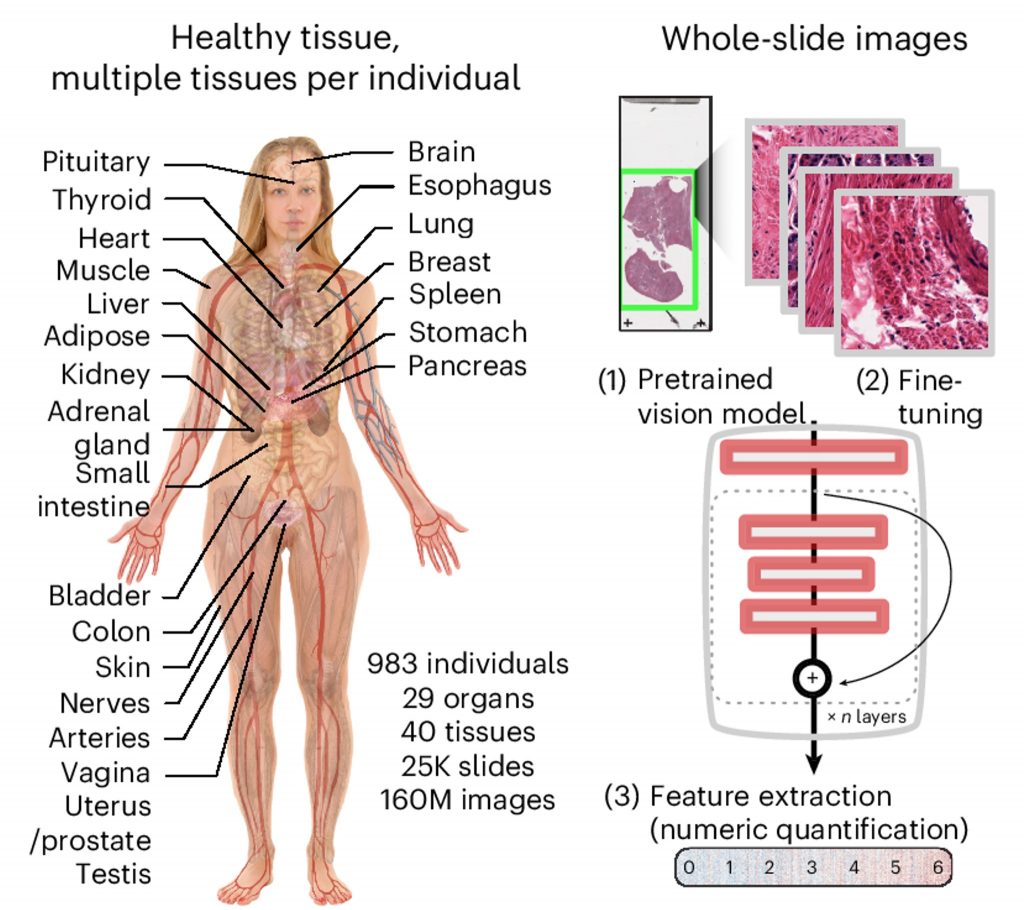
The researchers analyzed more than 25,000 tissue samples from the Genotype-Tissue Expression (GTEx) project, a resource that collects human tissues to investigate how genetic variation influences gene activity across the body. The study included tissue sections from 40 tissue types spanning 29 organs, collected from 983 people aged 20 to 70.
The researchers fine-tuned an AI vision model—a system designed to process and interpret images—to improve its accuracy. The model could detect structural features in tissue images and convert those differences into numerical measurements. Remarkably, aging emerged as the strongest driver of structural variation across tissues.
Age Gaps Are Associated with Chronic Diseases
Using the tissue samples, the researchers developed an “aging clock” that estimated age from tissue structure. They then compared each tissue’s predicted biological age with the donor’s actual age to calculate an age gap. Larger age gaps were associated with greater levels of pathology. For example, thickening of the aorta wall—a feature linked to cardiovascular disease—was associated with a larger age gap in aorta tissue.
The researchers found that age gaps in specific tissues were associated with chronic disease. Larger age gaps in lung tissue, for example, were linked to chronic respiratory disease, while larger gaps in pancreatic tissue were linked to type 2 diabetes. Age gaps in fat tissue were associated with heart disease, and those in prostate tissue were associated with high blood pressure. The strongest association was between kidney failure and age gaps across several tissues.
Common Age-Related Changes Across Organs
To better interpret age-related changes in tissue structure, the researchers used an AI model trained to connect tissue images with natural-language descriptions. This allowed them to assign aging-related terms to structural features in each organ.
Several features increased across multiple organs. This included fibrosis, the buildup of scar-like tissue that can impair an organ’s function; atrophy, or tissue shrinkage; and microvascular rarefaction, the loss or reduction of small blood vessels. Other features declined with age, including epithelium—the protective tissue that forms barriers and lines many surfaces—and hyperplasia, an increase in cell number that is often associated with tissue growth.
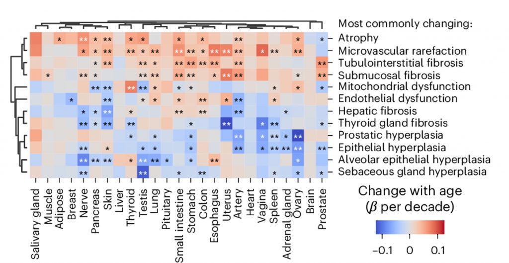
The researchers also found that tissue-based biological age aligned more closely with age-related changes in gene activity than the actual age of the donors. This may be because tissue architecture reflects the cumulative impact of many changes—including fibrosis, blood vessel loss, and shifts in cellular organization—that are not fully captured by measurements of cells in isolation.
Predicting Tissue-Specific Aging from Blood Samples
Having found that gene activity was linked to tissue age estimates, reflecting whole-body aging, the researchers sought to develop a tissue-specific age predictor. Using the gene-tissue link, they could measure gene activity from blood samples to derive age gaps in specific tissues. To validate their blood-based age predictor, they compared blood samples from healthy individuals to those of individuals with chronic diseases, such as Alzheimer’s disease.
They found significant correspondence between diseases and tissue-specific age gaps in at least one organ. For example, people with stroke showed age gaps specifically in the brain, kidney, and liver.
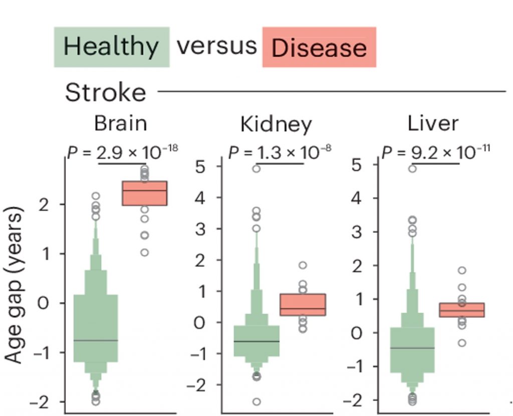
“Overall, this approach demonstrates the feasibility of inferring tissue-specific biological age and disease-related aging signatures from minimally invasive blood samples, underscoring its potential utility in early disease detection and monitoring aging in clinical settings,” said the authors of the study.
A Future of Precision Medicine
The findings of the Viennese researchers reframe aging as more than a collection of molecular changes within individual cells. By capturing how tissue structure changes over time—and linking those changes to gene activity in blood—the approach may offer a more complete view of how aging unfolds across the body. Although further validation will be needed before it can be used clinically, this kind of tissue-specific aging measure could eventually help identify organs under unusual strain, monitor disease earlier, and guide therapies toward the tissues most in need of intervention

 Comments