


Key Points:
- Researchers found that mitochondrial transplantation reduced several signs of heart aging and improved heart function in a mouse model of cardiac aging.
- The treatment appeared to work by helping restore the process in which heart cells clear and recycle damaged mitochondria (a process known as mitophagy).
- Although the findings suggest the potential of mitochondrial transplantation as a means to treat age-related heart dysfunction, human safety, mitochondrial delivery methods, and efficacy remain to be determined.
As published in Aging Cell, scientists from Shanxi Medical University in China report data on an experimental treatment approach in which mitochondria were isolated from stem cells and injected into mice modeling cardiac aging (a technique called mitochondrial transplantation). Interestingly, they found that mitochondrial transplantation lowered several markers of heart aging and improved heart function. Moreover, the treatment appeared to help restore the rate and completeness with which cells break down damaged mitochondria (a process called mitophagy). Results from the scientists’ study suggest that mitochondrial transplantation could treat age-related heart dysfunction; however, further research is needed to confirm safety, mitochondrial delivery methods, and the procedure’s efficacy in humans.
As the aging population grows at an accelerated rate, the burden of cardiovascular aging-related diseases increases as well. Furthermore, cardiovascular diseases have become one of the main causes of death and disability among the elderly.
Current treatments for cardiovascular diseases can modestly slow disease progression. However, they do not directly alleviate heart cell functional decline. Thus, there is an urgent need to develop therapeutic strategies that address the core pathological processes behind cardiac aging.
Relatedly, recent research has demonstrated that the transplantation of functional, intact mitochondria into damaged cells can improve cell function and promote tissue repair. Nevertheless, whether transplanting functional mitochondria into the heart translates into cardiovascular functional recovery during aging has remained unclear. To establish whether mitochondrial transplantation can restore functional aspects of an aged heart, the China-based researchers tested the procedure in a mouse model of cardiac aging.
More Details on Mitochondrial Transplantation
Mitochondrial transplantation is an experimental technique in which healthy, functioning mitochondria are isolated and administered to damaged cells or tissues. The aim of the procedure is to restore cell energy production, since in most cells, mitochondria are the main source of usable cellular energy in the form of ATP molecules.
For their study, the China-based researchers isolated mitochondria from stem cells and injected them into the tail veins of mice modeling cardiac aging. They then analyzed the effects of this technique on aspects of markers of cardiac aging and aspects of heart function.
The Mouse Model of Cardiac Aging
Analyzing cardiac aging in naturally aged mice can be time-consuming, since mice typically live 2 to 3 years in a laboratory setting. To address this, the researchers used a chemotherapeutic agent, doxorubicin, to induce cardiac damage similar to that seen with aging in younger mice. Accordingly, doxorubicin can recapitulate hallmarks of cardiac aging, such as mitochondrial dysfunction in heart cells.
Mitochondrial Transplantation Reversed Signs of Heart Aging and Improved Heart Function in the Cardiac Aging Model
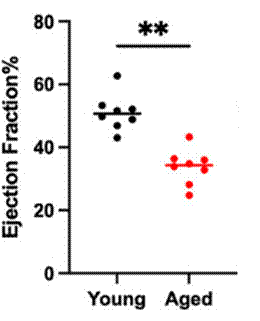
To confirm that doxorubicin indeed induces cardiac aging in young mice, the researchers measured aspects of cardiac function with echocardiography (an assessment that uses high-frequency sound waves to create moving images of the heart). In one of the echocardiogram measurements used, the ejection fraction percentage (the percentage of blood pumped out with each contraction of the heart), doxorubicin significantly reduced its average value. This finding shows that doxorubicin reduced the ejection fraction percentage, indicative of reduced pumping strength (a sign of cardiac aging that can increase the risk of heart disease).
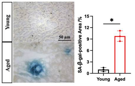
To further confirm doxorubicin’s induction of cardiac aging in cardiac tissue, the researchers analyzed levels of three protein markers of cellular senescence (a dysfunctional state of cells that occurs more frequently with advanced age). In cardiac tissue, all three of the cellular senescence markers were more than doubled. Collectively, these findings demonstrate that doxorubicin can induce aspects of cardiac aging in young mice.
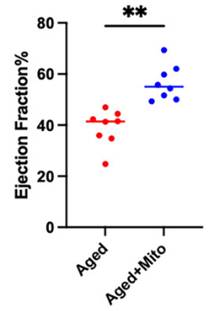
To find whether mitochondrial transplantation alleviates mitochondrial dysfunction and improves aspects of cardiac aging in this mouse model, the researchers injected stem cell-derived mitochondria into the mice modeling cardiac aging. They found that the transplantation significantly improved the heart ejection fraction percentage and normalized levels of protein markers of cellular senescence in cardiac tissue. Together, these results suggest that mitochondrial transplantation restores mitochondrial function and alleviates cardiac aging.
To uncover how mitochondrial transplantation may improve mitochondrial function in cardiac tissue, the researchers examined mitochondria under a microscope. They found that the mitochondrial transplantation technique restored the number of intact mitochondria in cardiac tissue and reduced the abnormal accumulation of vesicles formed around mitochondria selected for removal in mitophagy. These data suggest that mitochondrial transplantation increases the abundance of functional mitochondria in cardiac tissue by restoring mitophagy—the removal of dysfunctional mitochondria.
To uncover the cellular mechanism through which mitochondrial transplantation restores mitophagy, the researchers ran some experiments in mouse heart cells. Through multiple experiments, they identified a protein called HIF-3α that is sensitive to low ATP levels (as is seen with mitochondrial dysfunction during aging). HIF-3α acts to increase the levels of another protein called BNIP3 that induces the initiation of mitophagy but not the degradation of dysfunctional mitochondria. This scenario impedes mitophagy, leading to the congestion of dysfunctional mitochondria, which can induce senescence in heart cells and facilitate cardiac aging. Based on these findings, the researchers proposed that functional mitochondria from mitochondrial transplantation increase ATP levels to suppress HIF-3α and prevent cardiac aging.
A Need for Clinical Trials to Find Whether Mitochondrial Transplantation Prevents Cardiac Aging in Humans
Currently, no clinical trial has shown that mitochondrial transplantation reverses or treats cardiovascular aging in humans. Thus, the rejuvenative effects on the heart observed in this study cannot yet be extrapolated to older people with age-related heart dysfunction.
The limited human trial evidence available has focused on acute ischemia-reperfusion injury (the sudden tissue damage that can occur when blood flow returns after a period of low oxygen, such as after certain cardiac operations). More specifically regarding this evidence, mitochondrial transplantation has shown preliminary feasibility in small studies of acute cardiac injury. Still, more clinical research will be necessary to establish whether the technique is safe for humans; whether mitochondria from simple injections, as done in this study, survive enzymatic degradation in human circulation; and how effective the procedure is.
Along these lines, for cardiac aging, mitochondrial transplantation is unlikely to be broadly available as a standard human treatment soon. A realistic best-case estimate is that it could be available in 10 to 15 years, and it could take longer or never occur for treating cardiac aging. This is because there are no planned clinical trials in older adults that may show that it is safe and clinically beneficial. Thus, any aging research group interested in initiating such trials could surge to the forefront of the longevity research field by starting them in an effort to counter cardiovascular aging.
 Comments