


Key Points:
- Age was not associated with a steady decline in testosterone.
- Gold-standard body fat measurements (DXA scan) more strongly explained changes in testosterone.
- Waist size, an accessible and practical measurement, captured testosterone levels just as well as DXA.
- Maintaining testosterone levels with age may support a longer life in good health.
Low testosterone is often viewed as an inevitable consequence of growing older, encouraging people to seek testosterone replacement therapy (TRT). However, a new study published in the Journal of the Endocrine Society suggests that fat accumulation contributes to low testosterone more than age.
Researchers Dr. Adam Potter and Dr. Karl Friedl of the U.S. Army Research Institute of Environmental Medicine analyzed data from nearly 5,000 American men. They found that total body fat, fat surrounding the abdominal organs, and waist circumference were much more strongly associated with testosterone levels than age. Their findings suggest that evaluating one’s belly fat should take priority over simply attributing low testosterone to getting older.
Age Is Not Associated with a Steady Decline in Testosterone
The researchers examined data from 4,984 men ages 20 to 59 who participated in the National Health and Nutrition Examination Survey (NHANES) between 2011 and 2016. Alongside testosterone measurements, the researchers examined waist circumference and measured body fat using whole-body DXA (dual-energy X-ray absorptiometry) scans. The DXA scans were used to assess total body fat percentage and visceral fat, the fat stored deep inside the abdomen around internal organs.
Surprisingly, average testosterone did not decrease steadily across age groups. While men in their 20s averaged about 470 nanograms per deciliter (ng/dL) and men in their 40s averaged 400 ng/dL, men in their 50s averaged 410 ng/dL. However, the researchers compared different men rather than following the same men over time, so these results do not show whether an individual’s testosterone declines or rebounds with age.
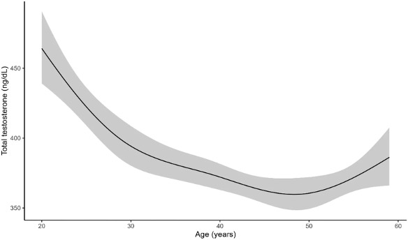
Age alone scored just 2.5% on a statistical measure of how well their models explained testosterone levels. In contrast, models combining age with total body fat, visceral fat, or waist circumference scored roughly 18–20%. These higher scores suggest that body fat and waist size provide substantially more explanatory power than age alone.
DXA-Scan Measurements Explain Changes in Testosterone Better than Age
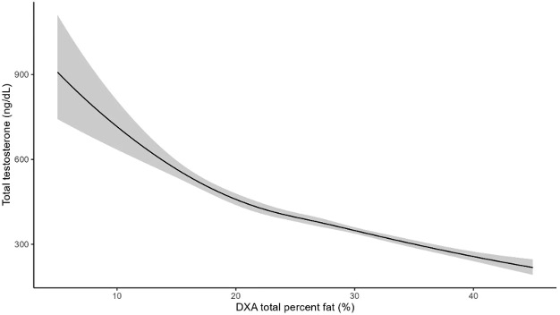
DXA scans are considered the gold standard for measuring body composition and fat levels. Based on the DXA scan measurements, the researchers’ age-adjusted models showed progressively lower testosterone levels as body fat percentage increased. For example, at approximately 15% body fat, predicted testosterone levels were nearly 600 ng/dL, while they dropped to about 400 ng/dL at 30% body fat.
The visceral fat measurements revealed a slightly more pronounced pattern, and once the researchers accounted for visceral fat, age was no longer consistently associated with testosterone levels. These findings suggest that fat accumulation helps explain differences in testosterone that were initially attributed to aging.
Waist Size Captures the Same Information as DXA Scan
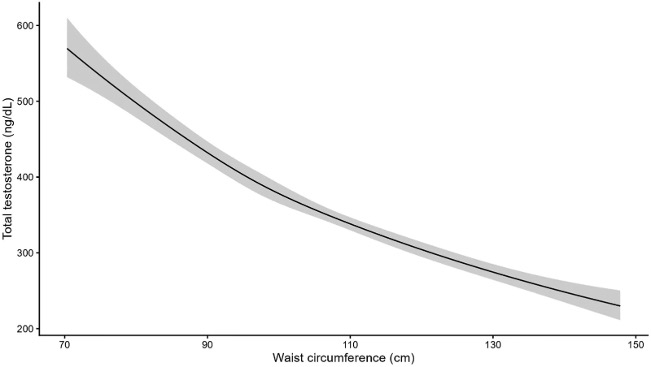
One particularly practical finding concerns waist size. The model incorporating waist circumference performed slightly better than the model incorporating DXA scan-measured visceral fat, scoring 19.9% versus 18.4% on the researchers’ measure of explanatory power. Although the difference was small, the findings suggest that a simple, inexpensive waist measurement can provide much of the same information about testosterone levels as specialized imaging.
Men With Low Testosterone Had More Fat, Not Less Lean Tissue
When the researchers compared men within the same age group, those with testosterone below 300 ng/dL (the cut-off used for low testosterone) consistently had higher body fat percentages and more visceral fat. Among men in their 20s, for example, those below the threshold averaged about 31% body fat, compared with 24% among those above it.
Interestingly, men with low testosterone also had a higher fat-free mass index, a measure of lean tissue relative to height, across every age group. Their larger bodies contained more lean tissue, but fat made up a greater share of their overall weight. This distinction suggests that low testosterone was associated more closely with excess fat than with a shortage of lean tissue, like muscle.
The findings also held when the researchers used stricter definitions of low testosterone—below 250, 200, or 150 ng/dL—rather than the original 300 ng/dL cutoff. The relationship therefore did not depend on choosing one particular threshold. These classifications were based on a single blood measurement, however, and do not establish a clinical diagnosis of testosterone deficiency.
Study Limitations
In addition to the single testosterone measurement, which may not accurately reflect each participant’s average testosterone levels, the study’s central limitation is that it demonstrates association, not cause and effect. It cannot establish whether excess fat lowered testosterone, or if low testosterone promoted fat accumulation. Moreover, other factors, such as medications, physical activity, and chronic disease, could influence both. Crucially, the cut-off age was 59, limiting interpretation for older adults.
What the Findings Mean for Health and Longevity
For health and longevity, the goal is not simply to raise testosterone, but to understand what a low reading may reveal about the body. Testosterone deficiency can affect sexual function, muscle, and bone health, and TRT can help appropriately diagnosed men—but it is not an established treatment for extending life or slowing aging. Excess visceral fat, meanwhile, is associated with diabetes, cardiovascular disease, and a higher risk of early death, making it a concern beyond its relationship with hormones. When excess fat accompanies low testosterone, losing the fat may improve both metabolic health and the body’s own testosterone production.
 Comments